
Case contribution: Dr. Radhiana Hassan
Clinical:
- A 17 years old male
- Known case of epilepsy with growth and pubertal delay
- History of breakthrough seizure due to non-compliance to medication
- Clinically: no neurological deficit
- EEG normal, blood investigation normal
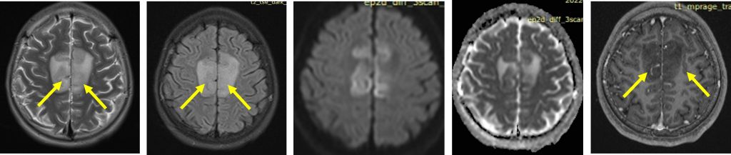
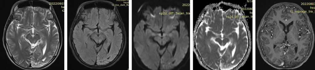
MRI findings:
- Abnormal thick cortex at parasagittal region of frontal lobe bilaterally
- It is isointense on T1, hyperintense on T2/FLAIR and no enhancement post contrast
- Minimal area of restricted diffusion also noted of the affected gyri
- No mass effect, no midline shift
- No abnormal vasculature
- An area of encephalomalacia also noted at left occipital region
Diagnosis: Polymicrogyria
Discussion:
- Polymicrogyria is also referred as cortical dysplasia
- It is a malformation due to abnormality in late neuronal migration and cortical organization.
- The neurons reached the cortex but abnormal with multiple small undulating gyri. It often appears fused on gross pathology and imaging giving rise to false impression of several large, thick gyri
- It has predilection for perisylvian region, when bilateral often syndromic
- CECT-difficult to detect the small convolutions of gyri due to poor resolution
- MRI shows irregular cortical surface, may appear as thick cortex with irregular gray-white matter junction without normal sulci or may appear as deep infolding or irregular thick cortex.
- Post contrast may show dysplastic meningeal veins overlying abnormal cortex.
Recent Comments