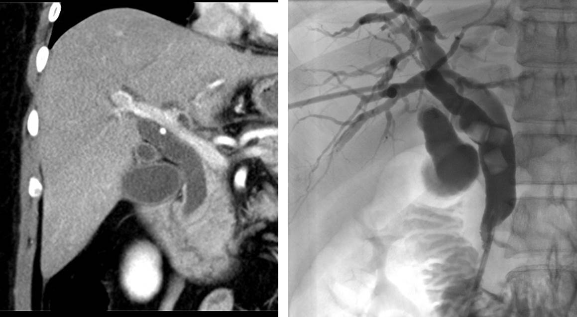
Radiolucent choledocholithiasis
Case contribution: Dr Radhiana Hassan Clinical: A 33 years old lady No known medical illness Presented with jaundice 2 days associated with pale stool and tea coloured urine. NO fever…
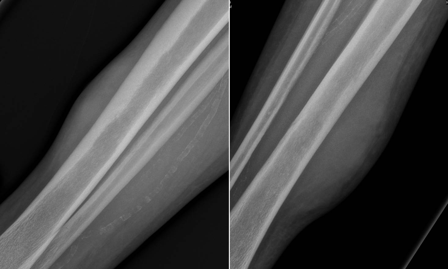
Primary lymphoma of the soft tissue (leg)
Clinical: A 73 years old man Underlying HPT and gouty arthritis Presented with mass at right leg for 5 months Initially started with redness at medial aspect of right leg,…
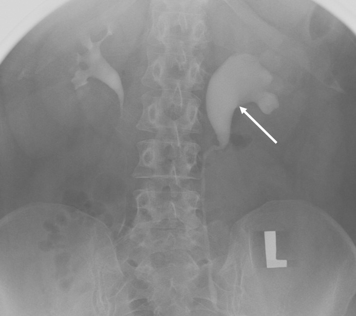
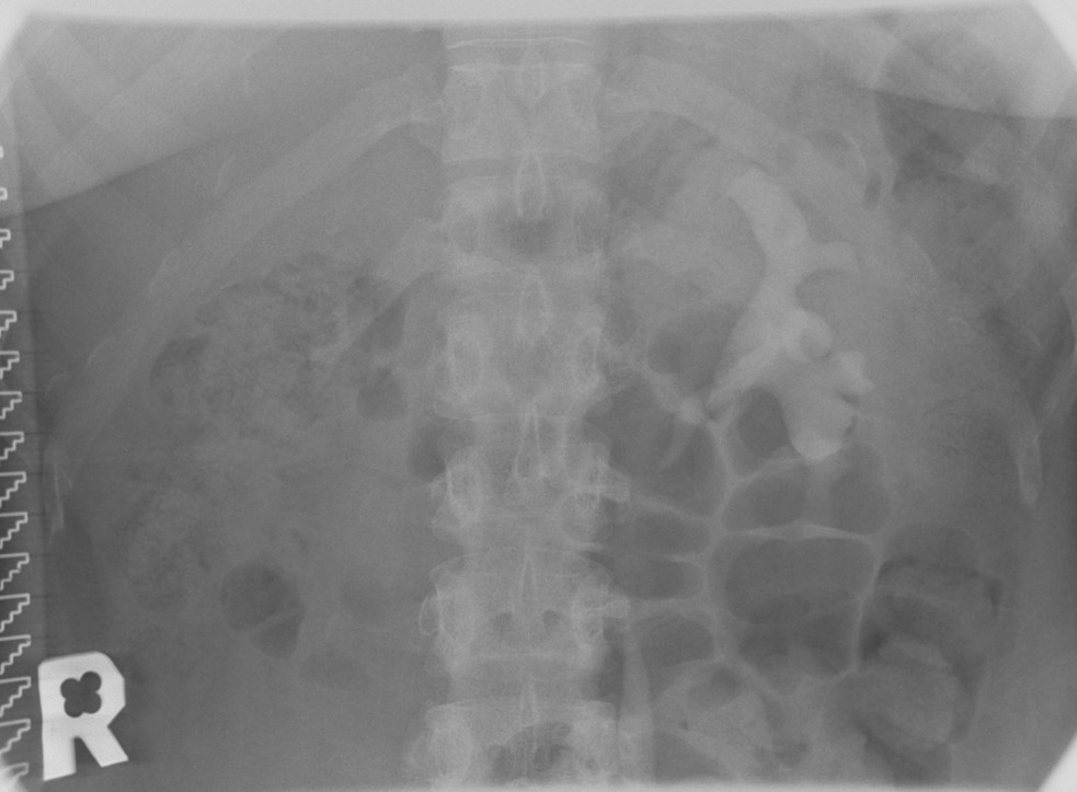
Pelviureteric junction obstruction
Clinical: A 51 years old lady History of recurrent flank pain No hematuria Imaging findings: Intravenous urography done No radiopaque calculus in preliminary film Normal right pelvicalyceal system and ureter….
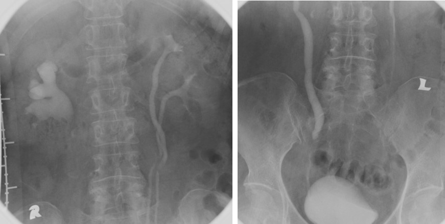
Adult ureteral stricture and duplex system of contralateral side
Clinical: A 64 years old lady Underlying DM and HPT Complaint or recurrent right flank pain Noted right hydronephrosis on ultrasound KUB For further assessment Imaging findings: Intravenous urography was…
Unilateral renal agenesis
Clinical: A 27 years old lady No medical illness Noted absent of right kidney on ultrasound Imaging findings: Intravenous urography perfomed. Normal opacification and demonstration of left renal and ureter…
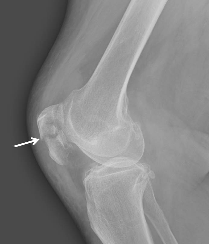
Patella fracture
Clinical: A 74 years old lady Alleged fall at home Complaint of painful swelling at right knee Radiographic findings: There is transverse fracture line at mid right patella (white arrow)….
Supracondylar fracture
Clinical: A 7 years old boy alleged fall and left elbow pain. TRO left supracondylar fracture. Radiographic findings: There is supracondylar fracture with subtle fracture line seen (yellow arrows) The…
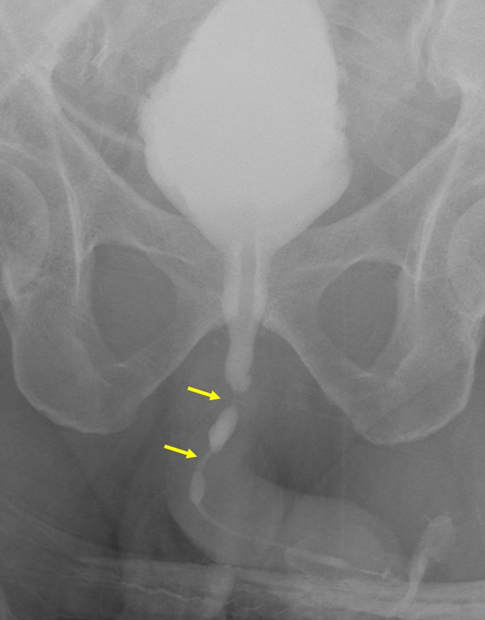
Anterior urethral strictures
Clinical: A 55 years old man History of Benign prostatic hyperplasia Recurrent acute urinary retention with history of CBD insertion Hematuria and pain after last CBD insertion Imaging findings: Ascending…
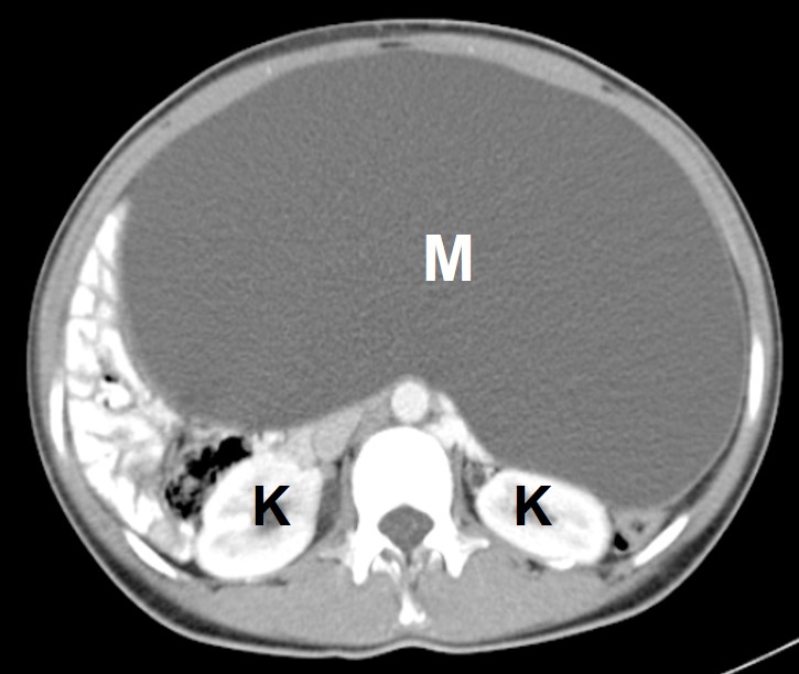
Ovarian serous cystadenoma
Clinical: A 24 years old lady Presented with progressive abdominal distension No constitutional symptoms CT scan findings: A large cystic mass arising from pelvic region measuring 10x23x34 cm. No calcification,…
Ovarian mucinous cystadenoma and uterine leiomyoma
Clinical: A 40 years old lady Presented with progressive abdominal distension No constitutional symptoms No bowel or urinary symptoms CT scan findings: A large non-enhancing cystic lesion arising from…
